
Congratulations, your loved one is pregnant!
Though it is too early for a pregnancy test to show what may be a long-awaited positive result, the embryo has already started to develop [1].
The blastocyst prepares for implantation into the uterine wall, and the mucous membrane releases tiny chorionic villi, the beginnings of the future placenta, to help it attach. The villi capture the blastocyst, spread the uterine tissue, and lead the way to the endometrium.
After implantation, the blastocyst begins to produce the pregnancy hormone, chorionic gonadotropin (hCG). The presence and level of hCG in the blood or urine serve to determine gestational age.
The inner and outer parts of the embryo start to form. The outer, or trophoblast, is responsible for the implantation of the embryo in the uterus. The internal, or embryoblast, assists in the development of the baby’s tissues and organs.
Between the inner and outer parts of the embryo, a liquid-filled bubble slowly forms. Surrounded by the chorionic villi, this bubble will become the placental barrier that protects the fetus.
What we can see on an ultrasound
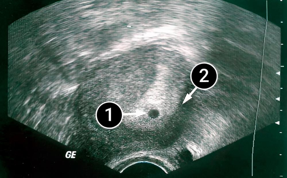
In the center of the picture, you can see a small dark dot, indicating a pregnancy with a single fetus. A thick layer of endometrium tightly surrounds the fetal sac. Where it meets the uterine wall, a vasculature and placenta will soon begin to form.
In the picture, the uterus is pear-shaped. At this time, the uterus has not started growing, and the mother is not yet showing.