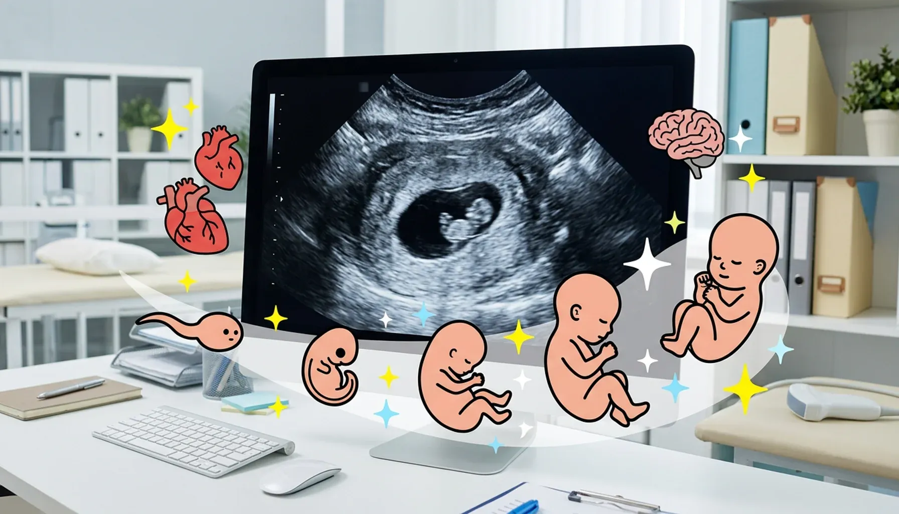

Your baby is growing!
On a three-dimensional ultrasound, the embryo looks like a tadpole. A pregnancy test will now show the much awaited positive result!

Your baby’s vital organs begin to form and he or she starts to receive nutrients from your blood supply. The blood delivers food and oxygen and also carries away waste — this is the beginning of uteroplacental circulation [1].
The placenta is beginning its incredible job of providing for your developing baby. While the baby develops, the placenta will enable she or he to:
breathe;
receive nutrients;
circulate blood;
produce hormones;
and suppress your immune system to a certain degree, to prevent it from mistakenly considering the fetus a foreign organism that needs to be attacked.
During this week, a strip appears on the surface of the embryo, which divides it in two. This is the axis of symmetry of the baby's developing body. The head and tail ends of the embryo are differentiated, the abdominal and dorsal surfaces are determined, and the spinal cord begins to form. The embryo folds in a longitudinal direction, taking a curved shape. At this stage, the embryo also becomes connected to the yolk sac. The site of connection will grow into the umbilical cord.
This week the nervous system begins to form and the foundations for the growing skin, muscles, bones, connective tissue, blood vessels and heart are laid.
The beginnings of the internal organs also appear this week. Almost simultaneously, the heart and genital glands of the fetus are created, and a little later the rudiments of the liver, lungs, trachea, intestines, pancreas and primary kidney are formed. In a male fetus, the sex glands are formed (the testes). Later, they will begin synthesizing testosterone — the main male sex hormone, which will ensure the further development of the fetus as a male type. In the absence of testosterone, the embryo will continue to develop as a female body.
What we can see on an ultrasound
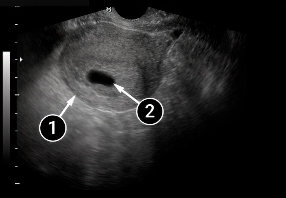
In the picture below, the rounded uterus is clearly visible. Inside it you can see the amniotic sac surrounded by the endometrium. The amniotic sac is the dark oval with clear contours located at the bottom of the uterus, which is considered one of the optimal positions for the sac.
At the current stage of pregnancy, the amniotic sac is still very small, only 5-7 mm.
The following photo shows a lengthwise view of a pear-shaped uterus. Within it, you can see a small black oval — this is the amniotic sac. It is surrounded by the endometrium, shown as the lighter outline. The lines of the inner walls of the uterus form the same pear-shape creating a cozy nest for baby.
uterus
amniotic sac
In this picture, you can also see the uterus and amniotic sac surrounded by the endometrium.
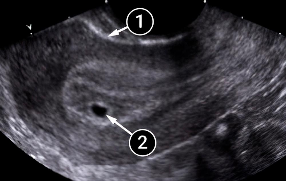
uterus
amniotic sac
In this picture, the embryo is visible! It’s the small white dot located in the amniotic sac. Both food and blood are supplied to the fetus by the endometrium.
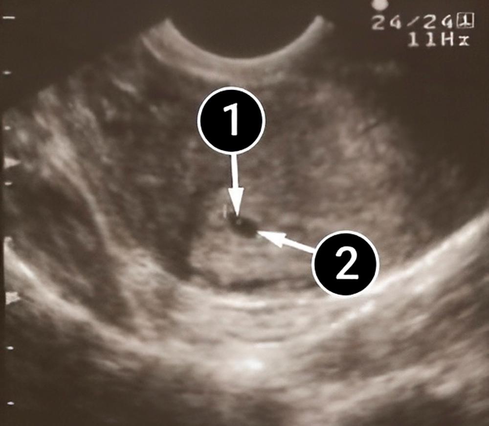
the embryo
amniotic sac
Brewer S. The Pregnant Body Book. Dorling Kindersley Publishing Staff, 2012, p. 87, 98, 102.