

Demystifying Conception
When a mature egg is released from a follicle, it is swept into the nearest fallopian tube. The fallopian tube, in turn, propels it towards the uterus. Fertilization usually occurs in the widest part of the fallopian tube, called the ampoule [1]. With intercours, millions of male sex gametes (or sperm) surround the egg, vying to fertilize it. Only one of them will penetrate the oocyte membrane (the scientific name for the egg) and merge with it, to create a new life.

As you may remember from high school biology class, the nuclei of the germ cells from the mother and father each contain 23 chromosomes. When the two come together, a full-fledged embryonic cell is formed — a zygote with a complete set of 46 chromosomes.
From the moment of conception, the zygote begins to divide to create new cells. However, at this point the embryo does not increase in size. By the end of the third day after fertilization, a morula forms from the zygote—this is the early stage of embryonic development. The morula looks like a blackberry and consists of 12 to 30 identical cells. Sometimes at this stage, for unexplained reasons, the zygote (fertilized egg cell) can split, resulting in two embryos, which will form into identical twins, also known as monozygotic twins.
When the morula reaches the uterus, it continues to develop for 3-4 more days and then becomes a blastocyst, which is implanted in the uterine lining. During the implantation period, the embryo is nourished by endometrial cells and will grow to be 0.2 mm in diameter. The implantation period lasts about 40 hours.
Meanwhile, where the egg was released from the follicle in the ovary, the corpus luteum begins to form, which consists of lutein cells that produce progesterone. This period of the menstrual cycle is called the luteal phase or the corpus luteum phase. In the absence of pregnancy, the corpus luteum degrades after 10-12 days and menstruation occurs. In the case of fertilization, it continues to develop. Until a placenta is formed, the corpus luteum is responsible for nourishing the new life growing inside you.
What can be seen on the ultrasound
At this stage of development, the future baby is called an embryo and is comprised of approximately 250 cells.
In the picture, the tiny white dot is the embryo, which is forming inside the fetal egg.
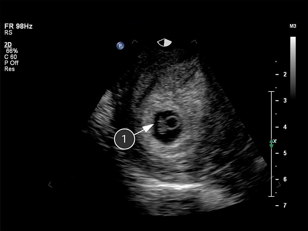
embryo
Brewer S. The Pregnant Body Book. Dorling Kindersley Publishing Staff, 2012, pp. 72-91.